
Medical imaging: There are various types of x-rays used for prevention and diagnostic purposes regarding oral diseases: extraoral and intraoral. A panoramic x-ray provides a broad vision of the upper and lower jawbone, nasal area, teeth, and temporomandibular joints.
In addition, the cephalometric images show teeth position in relation to the upper and lower jaw. These 3D images can create three-dimensional screenings of the internal structure.
Myray is one of the largest companies in the world in the production of devices to create medical and dental images. This company presents innovative solutions, which enable not only the success of the treatments, but also the improvement of people’s quality of life when they look for radiological solutions. They are at the forefront of technological development and operate in over 170 countries with about 800 patents.
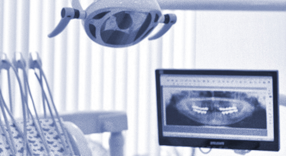
Panoramic system -Medical imaging

This panoramic system enables fast and effective diagnosis.
The communication with patients is clear and accurate. This device provides a complete and ideal diagnosis due to its high-quality panoramic, cephalometric and 3D images.
These high-quality images are extremely accurate and essential to dental implant treatment. In addition, the interface and software are easy to use, therefore, the exams will be simpler and quicker.
It is possible to select the 2D mode for traditional, traditional exams or, the 3D option can be used for more complex interventions, such as implantology or endodontics.
Advantages of the medical imaging – 3D Panoramic System
- It provides a complete overview of the teeth and upper and lower jaws, which is anatomically accurate and more detailed.
- It saves time and improves the treatment.
- It ensures a better image quality.
- The design of the device is easily adaptable to the patient, set according to the maxilla morphology with Comfort.
The Carestream Intraoral X-ray System
The intraoral x-ray, as it name suggests, enables the dentist to observe tooth decay and the tissues surrounding the tooth and its state. This is the most used x-ray in dentistry.
In our dental clinics abroad, we use various types of intraoral x-rays. We highlight the Carestream brand with high-frequency technology. This well-known brand was specially created for dental professionals and provides high-quality results, which makes the diagnosis more accurate and correct. It has a smart and flexible design to meet the demands of the user.
Advantages of Intraoral X-Ray
- Accurate images that are easily acquired.
- It is adaptable to almost all imaging technology.
- The diagnosis is safe, accurate, and consistent.
- Thanks to the new design, it is possible to get a correct alignment between the head of the x-ray tube, reducing the risk of cut images.
RVG Dürr Dental
The company Dürr Dental AG was established more than 70 years ago in Germany. This company provides various solutions in several fields of dentistry, such as equipment and diagnosis systems. It is present in more than 36 countries in Europe and across the world, employing over 1000 workers and with sales over 200 million euros, which reflects its business success. Dentists around the world trust in its engineering and continuous development. This medical and technological company was considered one of the best SMEs in Germany several times, winning numerous awards and distinctions.
RVG stands for intraoral digital radiology or radio-videography. This RVG device provides intraoral radiographic and digital images. It provides high-definition and high-contrast images and, for that reason, it makes the diagnosis easier. Its sensor is sophisticated, very easy to use, and compatible with most of the radiological and management software of a dental clinic. The users can obtain, visualise, adjust, and save the images on their iPods or iPhone and later transfer them to a computer to analyse or just store the content.
Through the use of this device, it is possible to visualise images with small dimensions, which is essential in dentistry.
Intraoral Camera – medical imaging
The intraoral digital camera helps to identify emerging cavities in teeth during a screen exploration. After identifying the cavity, the dentist can save the image and analyse it with great detail.
These cameras consist of small, high-power LEDs with a spherical lens and a condenser that lights the area, providing sharp images with extraordinary luminosity.
Diagnosis and Planning – Digital Solutions
Through the use of NobelClinician, it is possible to accurately plan supported-implant solutions. This software is considered the newest, most modern and complex technology for the diagnosis and digital planning of clinical cases. Thanks to its virtual environment, 3D radiographic information of patients can be studied and analysed in detail, from its initial stage to the prosthetic stage.
NobelCliniciam Software Advantages
-
More accurate diagnosis:
It is possible to simultaneously visualise various areas that may need intervention. This visualisation with panorex, transversal, and 3D views enables a significant increase in the accuracy and quality of the clinical diagnosis.
-
Treatment Plan is more effective:
From an anatomical perspective, the bone model creates the possibility to visualize and accurately define where the titanium rods will be inserted. In addition, the radiological guide will provide virtual images of the teeth. Through these images, dentists will be able to find a better position for the teeth and titanium rods in order to achieve a better oral rehabilitation.
-
The information for the patient is better:
The dentist and the candidate for oral rehabilitation can discuss the treatment plan. Various treatment options will be explained and analysed.