Methods for Diagnosis Purposes

These methods contribute to the development of dentistry, particularly in implantology. The technical advances of this dentistry speciality were possible thanks to collaboration on diagnosis methods.
In addition to a careful clinical exam carried out by the dentist, x-ray analysis enables the planning of an accurate diagnosis and success in cases of implant rehabilitation and aesthetical restorations.
The x-ray images taken from different angles produce multiple cross-sectional images that can be simple or bi-dimensional. The radiology methods applied to oral implantology are called orthopantomogram, intraoral radiography, and computerised axial tomography scan (CAT scan).
What is orthopantomogram?
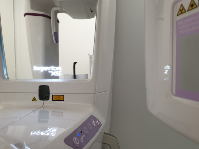
Orthopantomogram is commonly known as a panoramic x-ray and provides two-dimensional maxilla and mandible images.
This procedure provides important information regarding height, bone width, and the necessary bone in the areas for implant rehabilitation. Through the use of a panoramic x-ray, the dentist will decide on the best location and angle to place the titanium rods.
What is the aim of the orthopantomogram two-dimensional images?
With these two-dimensional images, the dentist will see all the alveolar-dental structures and analyse various characteristics, such as the maxillary sinuses, nasal passages, inferior dental nerve route, and the locations of the mental nerve holes. It is possible to make accurate vertical measures. As the field of view is wide, intraoral x-rays may be necessary. In other words, another radiology method should be considered. Subsequently, intraoral x-rays should be taken to visualise other details that may not be visible in the panoramic x-rays.
What type of pathologies can the panoramic x-ray identify?
- Pathologic changes, congenital conditions, or conditions resulting from other factors.
- Tooth decay.
- Pulp disorders.
- Periapical pathologies.
- Gum diseases.
Disadvantages of a panoramic x-ray
The images are only two-dimensional and the target area is almost always three-dimensional. It does not provide information in a transversal angle, in other words, information regarding bone density.
Most equipment has positioning guides, but sometimes patients may be incorrectly positioned. This can cause errors, such as tongue structures being displayed in a much higher position than the vestibular structures.
Computerised Axial Tomography Scan (CAT scan)
What is CAT?
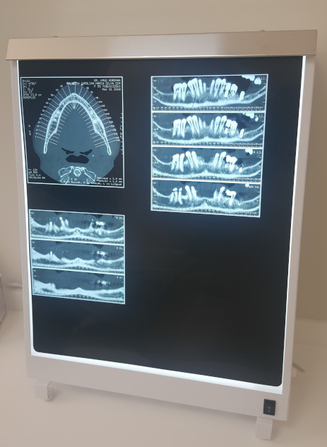
CAT scan, or computerised axial tomography scan, provides three-dimensional images. It makes use of computer-processed combinations of many x-ray images taken from different maxillary angles to produce cross-sectional and panoramic images of maxillary areas. Cross-sectional images can also be obtained. CAT scans will provide high-definition images and those measurements correspond to the real size.
Advantages of CAT scans

They provide homogenous magnification with high-definition and high-contrast images.
These x-rays enable better structure identification of bone grafting used in sinus lifts. In contrast, a panoramic x-ray only has two-dimensional images, which makes it difficult to identify these structures. The images from a computerised axial tomography scan do not have any distortions or overlaps.
The specialised software enables analysis of the obtained images.
In this way, it is possible to obtain information regarding height, width, alveolar structure inclination, and also the anatomical and topographical structures. There are images from the maxilla and mandible anomalies as well as the location of the inferior dental nerve, sinus, among others.
There is no need to apply a correction coefficient as the axial images with 1 mm are printed in real size together with a scale to measure them. The user can immediately measure the distance between different images.
The importance of CAT scans in Implantology
There are various software programs that enable pre-simulation, with two-dimensional or three-dimensional images, of the process involving implant placement. This software optimises the entire process and decreases the possibility of errors during planning.
Disadvantages of CAT scans
The greatest disadvantage is the high cost of the equipment, which also makes the exam quite costly. The radiation dosage is slightly higher in comparison with other radiology methods. Despite the higher radiation and cost, this method has so many benefits that its use is completely reasonable for implant rehabilitation.
Conclusion About Radiology
 Panoramic x-rays are an excellent diagnostic method and is utilized for specific areas as it provides two-dimensional images. It has lower doses of radiation, which is a great advantage. It is a standard exam for implant surgeries and planning of the case. The risk of radiation is minimal and it is able to accurately determine the length of the implant that will be placed.
Intraoral x-rays are used to complement images obtained from panoramic x-rays in areas that may raise doubts due to poor image definition.
Cut-sectional images should be taken in special cases of implantology and when required by the dentist. If the health professional detects bone loss or requires more information about the sinus structure, he/she will require this method. It is highly recommended for edentulous persons and is mainly used for complex surgical procedures.
Reviewed by VitaCentre Dental Clinic Staff on June 1, 2023

